Abstract
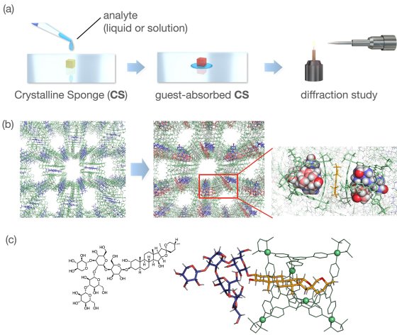
Among various molecular structure analysis methods, single crystal diffraction stands out as the most reliable, providing precise three-dimensional structures of molecules. In 2013, our group introduced a new X-ray structure analysis method termed the crystalline sponge (CS) method, eliminating the need for analyte crystallization. In this method, a small crystal of a porous complex (CS) is immersed in a solution containing the target compound, which is then absorbed and aligned within the CS pores. The post-oriented (or "post- crystallized") target compound can then be observed alongside the host CS framework through crystallographic analysis. Despite its innovation, the CS method has certain limitations related to, for example, molecular size and polarity of analytes. Here, an M6L4 cage, developed over the past three decades with extensive host-guest chemistry, is employed as a potent CS to address the several limitations of the original method. Large aromatic polysulfonates ("sticker" anions) significantly facilitated the crystallization to give cage-stacked single-crystalline sponge (sc-1) . The symmetry mismatch between the cage (Td) and the sticker (D2h) resulted in a low space group (typically P1), avoiding the static guest disorder problem and leaving guest-accessible channels in the crystal. Guest accommodation in the cage cavity can be performed either before or after the cage crystallization. Thanks to the cage's large cavity with high guest-binding properties, a broader range of analytes can be analyzed, including water-soluble molecules, large amphiphilic molecules (MW ~1200), and molecular aggregates prior to reactions (Figure 2).

