CIRB - Équipe de recherche

Functioning and Adaptation of Microorganisms

Fonctionnement et adaptation des microorganismes

Principal Investigator: Olivier ESPELI, DR1 CNRS

Présentation

Our group studies the adaptation of bacteria to changes in their environments. Although bacteria are among the simplest living organisms, formed of a single cell, they display a huge adaptability to the presence of nutrients or toxic compounds in their environment. This adaptability allows them to survive periods of nutrient depletion, treatment with antibiotics or survival within their host in the context of an infection.  

The first line of adaptation goes through the modulation of gene expression. This characteristic is known for decades. Since the pioneer work of Francois Jacob, Jacques Monod and their colleagues on the lactose operon in 1961, this field has been revolutionized by the development of genomic methods. In the laboratory, we use cell biology and genomic assays to analyze the expression of genes in response to various stimuli, the binding of proteins to multiple sites on the bacterial chromosome and even the 3D folding of the chromosome in various growing conditions.

Figure 1: Coupling between gene expression (green), cell growth (blue) and chromosome folding (red) in bacteria

We observed that chromosome folding and genome expression are tightly connected (Lioy et al 2018, Planchenault et al 2020).We study various aspects of the interplay between chromosome folding and gene expression; we analyze of the role of the physics of the DNA molecule in transcription initiation and the role of transcription on long range DNA folding.

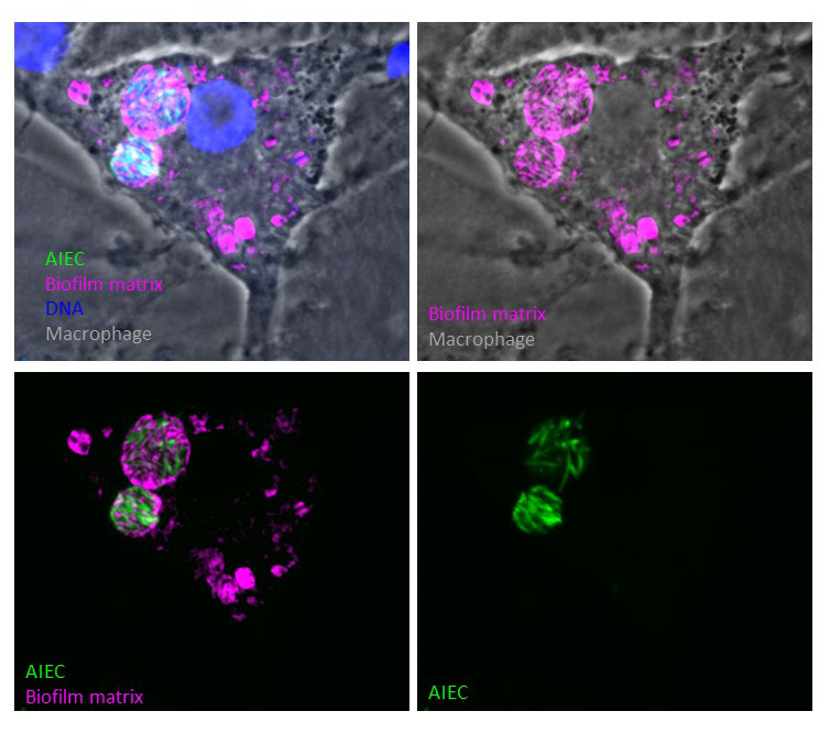
A second aspect of bacterial adaptation relies in modification of the growing regime and life style of the bacteria. In the presence of a stress bacteria can halt their cell cycle to limit the extent of the damages; in the harshest conditions, some bacteria may enter in a dormancy state, where metabolic activities are limited. This state confer them a high tolerance to antibiotic for example. In other conditions, bacteria cooperate to form communities, also called biofilm, that favor their adherence and tolerance to stresses.

Localisation of a genes in the bacterial cell (Espeli)
Figure 2: Localisation of genes (blue, green and red foci) in bacterial cells (gold)

In the recent years we have observed, with commensal Escherichia coli, that chemotherapeutic drugs such as Mitomycine C, Bleomycin or AZT severely change the cell cycle. This is linked to the sudden expression of dozen of genes from the SOS regulon. One protein from this regulon caught our attention, RecN. This protein belongs to the SMC family that also contains eukaryotic cohesins and condensins . When a particular type of DNA Damage repair is engaged, RecN is able to block chromosome segregation and to promote the regression of already segregated sister chromatids. RecN acts as a DNA damage specific cohesin, this is the first bacterial cohesin described (Vickridge et al 2017). In E. coli, sister chromatid cohesion is a transient step usually mediated by topological links between sister chromatids. Our group contributed to the discovery of this topological cohesion process (Lesterlin et al 2012) and characterized some aspect of its regulation (El Sayyed et al 2016). We studied the impact of the sister chromatid cohesion step on global chromosome folding in normal or pathological cell cycles (Conin et al 2022).

Movie 1: Impact of chemotherapeutic drugs on E. coli growth in the absence of RecN

The E. coli chromosome is labelled with the HU-mCherry fluorescent protein, mitomycine C is added for 5 minutes to the growing medium then washed. The total length of the experiment is 4 hours.
 

Our interest for bacterial adaptation to the environment brought us to study bacterial pathogens whose environment is shaped by their host. We work with E. coli, Shigella and Listeria that are intracellular pathogens. They can survive and multiply within macrophages or epithelial cells. Adherent Invasive E. coli (AIEC) have been identified in Crohn’s disease patients. They colonize the digestive tract, adhere to epithelium and multiply within macrophages that are supposed to eliminate bacterial pathogens. AIEC present an original colonization of macrophages, they do not escape from the phagolytic vacuole and they do not detoxify it, instead they multiply in a toxic environment (acidic, oxidative, nutrient poor). To do so they have to adapt permanently. We demonstrated that for AIEC LF82, this adaptation involves at least two steps (Demarre et al 2019). First, LF82 bacteria halt their cell cycle to become dormant for several hours. During this period, they change their gene expression program to construct a protective matrix, akin to a biofilm matrix. Once the matrix program is initiated, bacterial multiplication starts again and bacterial communities form (Prudent et al 2021). These intracellular bacterial communities can serve as a long-term survival niche for AIEC in a low pH, oxidative and toxic environment. In this frame, we are involved in the European Community COST action EuromicropH aiming at characterizing microorganisms’ life at low pH.

Figure 3: Human macrophages infected by AIEC bacteria (red). The bacteria form intracellular communities inside toxic phagolysosomes labeled by Lamp1 (green)
Figure 3: Human macrophages infected by AIEC bacteria (green). Inside phagolysosomes, the bacteria form intracellular communities surounded by a biofilm matrix (magenta).

To understand AIEC strategies during macrophage infection we explore different paths. First, we evaluate the relevance of the IBC way of life for different AIEC strains collected from Crohn’s disease patients. Second, we explore how these bacteria manipulate metal ions homeostasis during infection. Third, we are characterizing the cell cycle management (replication, segregation, repair of DNA, cytokinesis) of dormant persistant bacteria. Finally, we study dynamics and evolution at the single bacterium level of communities embedded in a biofilm matrix.

Movie 2: Expression of the AIEC LF82 pathogenicity island (green) allowing iron capture during infection of macrophages. 

Our projects are funded by The Agence Nationale de la Recherche , by the Labex Memolife from PSL university, the Qlife program (https://fr.qlife.psl.eu/), the ARC foundation, the Association Francois Aupetit (AFA), the Foundation Saint Michel du Collège de France, the Weizmann Foundation and the European Community COST action EuromicropH.

Équipe

Senior researcher:
Sylvie Rimsky, DR2 CNRS

Postdoctoral fellows, PhD students:
Céline Borde, Postdoctoral fellow
Stéphane Biedzinski, Postdoctoral fellow
Emma Bruder, PhD student
Hosni Nedjar, PhD student
Justine Groseille, PhD student

Technical staff:
Quenech’du Nicole, IEHC CDF
Lisa Bruno, IE Qlife program

Collaborators:
Olivier Rivoire (CIRB, Collège de France)
Romain Koszul (Pasteur Institute, Paris)
François Xavier Barre (I2BC, CNRS, Gif sur Yvette)
Nicolas Barnich (M2iSH, Université Clermont Auvergne, Clermont Ferrand)
Clotilde Policar (IBENS, ENS, Paris)
Silvia De Monte (IBENS, ENS,Paris)
Guy Tran Van Nhieu (I2BC, CNRS, Gif sur Yvette)
Marie-Agnès Petit (Micalis, INRAE, Jouy en Josas)
François Cornet (CBI, Université de Toulouse)
Ivan Junier (TIMC, Université de Grenoble)

Alumni:
Hafez El Sayyed (Oxford University, Oxford, UK)
Elise Vickridge (Mc Gill University, Montreal, Canada)
Gaëlle Demarre (Pherecydes Pharma)
Victoria Prudent (UCSF, San Franciso, USA)
Charlene Planchenault (Kelly Scientifique)
Charlotte Cockram (Oxford Biomedica, Oxford, UK)
Parul Singh (Institut Pasteur, Paris)
Brenna Conin (Synchrotron Soleil, Gif sur Yvette)