
A topical scientific issue explored by a researcher from the Collège de France.
Published on 22 October 2026
News
What causes an oocyte to grow?
Spotlight
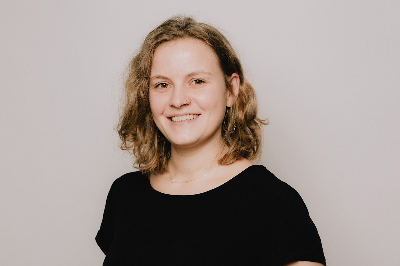
As they grow, oocytes – the female reproductive cells in mammals – accumulate the resources necessary for their future fertilisation and pre-implantation development. A recently discovered cluster in mouse oocytes appears precisely when this growth accelerates. Initial observations of this structure have led to a better understanding of oocyte development and could, in the long term, help to determine which oocytes are suitable for fertilisation and show good potential for future development.
We spoke to Noemi Zollo*, a biologist at the Collège de France.
Before it can be fertilised, the oocyte must grow and accumulate the reserves necessary for the early stages of embryonic development. However, once this growth is complete, assessing the quality of the oocyte remains a challenge. This difficulty arises, for example, during in vitro fertilisation, a technique used in assisted reproductive technology. Oocytes that have completed their growth and resumed cell division – and are therefore considered ready for fertilisation – are retrieved from the ovaries to be fertilised by sperm in the laboratory. “But the size of the oocytes alone is not enough to determine whether they are of good quality,” emphasises Noemi Zollo. Many studies have focused on the stages leading up to the formation of oocytes and on those – particularly cell division – that enable them to be fertilised. However, the growth of these cells between these two phases remains less well understood. The observation of an unexpected structure has opened up a new avenue for understanding what happens during this expansion.
Discovering the Zollo body
Noemi Zollo was due to study the final phase of mouse oocyte growth. But, out of curiosity, the PhD student also turned her attention to smaller cells. Using a marker that reveals RNA – molecules used, amongst other things, in the production of proteins – she noticed a condensed cluster near the nucleus. “To begin with, I had no experience with oocytes, so I didn’t know what was ‘normal’ or not,” she explains. On the advice of her thesis supervisors, Maria Almonacid and Marie-Hélène Verlhac, the researcher consulted the scientific literature to determine whether her discovery corresponded to a known structure or whether it warranted a study in its own right, and realised that it had never been described before. “I couldn’t believe it. I was just curious and I was lucky, because I wasn’t actually supposed to be studying this in the first place,” she continues.
At first, the biologist thought she had observed a cluster resembling the Balbiani body, a structure known to exist in the oocytes of many animals, but which appears earlier in their development. To investigate this similarity, the researcher used antibodies – proteins in the immune system capable of recognising a specific target and binding to it. By selecting those that target proteins characteristic of the Balbiani body, she hoped to detect them in this new component. “It was really frustrating , ” recalls Noemi Zollo, “because I couldn’t find the proteins. ’ She then changed her approach, so as no longer to limit her work to the proteins expected on the basis of a resemblance to the Balbiani body. Rather than searching for potential elements identified in advance, she extracted the clusters directly from the oocytes to isolate them. She then analysed them using mass spectrometry, a technique that makes it possible, amongst other things, to determine which proteins are present in a sample.
Named ‘Zollo body’ by Marie-Hélène Verlhac, in honour of the PhD student, the assembly comprises RNAs, proteins and several types of organelles – small structures, each of which performs a specific function. To understand the role of the Zollo body within the oocyte, the biologist examined the RNAs it contains in greater detail. Some carry the instructions needed to produce proteins, a process known as translation. Among these, she identifies RNAs that enable the production of proteins involved in the growth of the oocyte and the follicle – the structure that surrounds the oocyte in the ovary. Others are used to produce proteins that themselves play a part in this translation process. The cluster thus comprises RNAs likely to contribute to the cell’s growth and its ability to produce new proteins. The researcher also notes that the Zollo body appears when the oocyte reaches a diameter of around 60 micrometres. It takes approximately two weeks for the oocyte to reach this size from an initial size of 20 to 40 micrometres. “At this stage, growth accelerates dramatically: in just two to three days, the oocyte reaches its final size, around 80 micrometres,” explains Noemi Zollo.
Based on these observations, she hypothesises that the Zollo body plays an active role in the growth of the oocyte and its follicle. It is thought to promote the production of the proteins and organelles necessary for the cell’s development as it prepares for fertilisation.
A role in oocyte growth
“Simply observing the Zollo body whilst the oocyte is growing rapidly is not enough to prove that it contributes to this growth,” the researcher points out. To test her hypothesis, she first verified that the cell had accumulated proteins as it grew, rather than simply swelling by diluting its contents. She then compared protein concentrations at different stages of development and found that these remained stable despite the increase in the oocyte’s volume. The total quantity of proteins therefore increases in line with its size. These measurements establish that the cell accumulates proteins as it grows, but do not yet allow us to attribute this accumulation to the Zollo body. ‘The isolated clusters allowed us to analyse their composition. But to study their role in growth, we needed to track the oocyte along with the follicle surrounding it over several days , ’ explains Noemi Zollo. ‘So we observed them in ovarian tissue.’ In this preserved environment, she rediscovered the characteristic structure of her discovery.
The biologist moved on from observation to disrupting the assembly. “Among the proteins identified, we pinpointed DDX5, which may act as a glue, holding all the components together so they do not disperse within the oocyte,” she explains. She blocked its activity to examine the consequences for oocyte growth. “After inhibiting DDX5, the cluster broke up, and the growth of the oocyte and its follicle came to a complete halt!” reports Noemi Zollo. Control experiments have also confirmed that this halt cannot be explained simply by the experimental conditions. The active role of the assembly in the development of the cell and its follicle has thus been proven.
The team is now seeking to verify whether the proteins produced within the Zollo body do indeed contribute to the formation of new organelles. Noemi Zollo, for her part, would like to gain a better understanding of what maintains the assembly. She is particularly interested in the ‘glue’-like role attributed to DDX5. “This protein is relatively scarce within the structure, so perhaps other proteins could also play this role , ” explains the researcher. She also wishes to study the oocyte and the follicle surrounding it together, as they are currently often analysed separately.
As the experiments were carried out in mice, the presence of the Zollo body in humans remains to be established. However, access to oocytes at the right stage of development poses a major obstacle. Those donated to research following in vitro fertilisation treatment have often already completed their growth. “At this stage, the cluster would already have disappeared if it were following the same developmental path as in mice , ” explains Noemi Zollo. As for the oocytes that did not respond to treatment, the researcher is investigating the reasons for this lack of response and whether there might be a problem with their development. Research is continuing in mice, with a broader aim: the biologist wishes to understand whether all oocytes go through this stage to complete their growth and assemble all the components necessary for fertilisation. Beyond the discovery of a structure, the aim is to understand how a reproductive cell prepares to enable the beginning of a new life.
*Noemi Zollo holds a PhD in cell biology, having completed her doctoral research within the Oocyte Mechanics and Morphogenesis team at the Interdisciplinary Centre for Biological Research at the Collège de France, under the supervision of Drs Marie-Hélène Verlhac, Maria Almonacid and Gilles Tessier (Sorbonne University).


Latest associated news
All news
Take Action with the Collège de France Foundation
Take Action with the Collège de France Foundation
Published on 2 October 2026
Clinical Research Assistant (F/M)
Job vacancy
Clinical Research Assistant (F/M)
Published on 20 October 2026

Postdoctoral Researcher (F/M)
Job vacancy
Postdoctoral Researcher (F/M)
Published on 20 October 2026
Postdoctoral Researcher (F/M)
Job vacancy
Postdoctoral Researcher (F/M)
Published on 20 October 2026
2026 Nobel Prize in Chemistry
2026 Nobel Prize in Chemistry
Published on 9 October 2026
Atoms to Recreate the Quantum Chaos of Black Holes
Atoms to Recreate the Quantum Chaos of Black Holes
Published on 23 September 2026
Catherine Llorens-Cortes is the recipient of the 2026 Prize of Honor from the National Academy of Pharmacy
Catherine Llorens-Cortes is the recipient of the 2026 Prize of Honor from the National Academy of Pharmacy
Published on 14 September 2026

Postdoctoral Researcher (F/M), Chemistry of Biological Processes Laboratory – Prof. Fontecave
Job Opening
Postdoctoral Researcher (F/M), Chemistry of Biological Processes Laboratory – Prof. Fontecave
Published on 8 September 2026